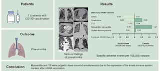
COVID-19 vaccine-related pneumonitis
COVID-19 vaccine-related pneumonitis (CV-P) should be considered in patients with atypical lung infiltration, no specific etiologies, and recent COVID-19 vaccination.
An article posted ahead of print by the Korean Journal of Internal Medicine describes 11 cases with a median age of 80. 10 individuals survived, but one patient with acute respiratory distress syndrome (ARDS), an 82-year-old-female, died despite appropriate treatment. Her chest CT displayed diffuse alveolar damage (DAD pattern).
Here are several other cases:
An 83-year-old woman who received the second dose of BNT162b2-mRNA vaccine presented with an organizing pneumonia pattern of CV-P. Chest CT revealed multifocal patchy consolidations, ground-glass opacifications (GGOs), and mild interlobar septal thickening in both lungs (14 days after the second dose). BAL fluid analysis revealed neutrophils (Np) 24%, lymphocytes (Lp) 32%, eosinophils (Eo) 32%, and macrophages (Mq) 12%. All microbiological tests in BAL specimens were negative. Intravenous methylprednisolone was initiated at 24 mg/day, and the patient had a rapid response to treatment.
73-year-old man who received the first dose of ChAdOx1 nCoV-19 vaccine. Chest CT revealed extensive bilateral GGOs with consolidations. There were no abnormal findings in the lung parenchyma on chest CT performed 10 months prior. BAL fluid analysis revealed Np 29%, Lp 32%, Eo 2%, and Mq 37%. The interstitial lesions gradually improved with steroid treatment.
51-year-old woman received the first dose of BNT162b2-mRNA vaccine. 8 days after vaccination, chest CT showed diffuse bilateral GGOs, centrilobular nodules, bronchovascular bundles, and interlobular septal thickening, but no cardiomegaly. Small bilateral pleural effusions were present, but not shown in the current figure. BAL fluid analysis revealed Np 0%, Lp 92%, Eo 7%, and Mq 1%. Methylprednisolone was initiated at 90 mg/day; rapid clinical and radiological improvement was observed.
An 80-year-old woman was diagnosed with HP following mold exposure 2 years prior. She changed her living accommodations and received corticosteroid therapy for 3 months resulting in complete remission and was relapse-free after discontinuation of steroids. Cough with sputum production developed 7 days after BNT162b2-mRNA vaccination, and chest CT showed recurrence of centrilobular nodules, GGOs, and a mosaic pattern of HP. She improved with steroid treatment.
Acute exacerbation of IPF was observed in an 80-year-old man who received BNT162b2-mRNA vaccine (first dose). He discontinued pirfenidone 1 year before vaccination due to adverse skin reactions and anorexia. Four days after COVID-19 vaccination, chest CT revealed new bilateral GGOs superimposed on reticular opacities and honeycombing with spontaneous pneumothorax. The patient improved with steroid treatment and chest tube management and was discharged with no acute exacerbation thereafter.
REFERENCE
Ji Young Park, Joo-Hee Kim, Sunghoon Park, Yong Il Hwang, Hwan Il Kim, Seung Hun Jang, Ki-Suck Jung, Yong Kyun Kim, Hyun Ah Kim, and In Jae Lee. Clinical characteristics of patients with COVID-19 vaccine-related pneumonitis: a case series and literature review. Korean J Intern Med 2022/08/22 doi: 10.3904/kjim.2022.072


Comments
Post a Comment