A
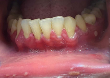
38‐year‐old Thai woman developed painful oral lesions lasting over 8 weeks. The lesions appeared 1 week following the administration of the first dose of AZD1222 vaccine.
Her extraoral examination was unremarkable. The intraoral examination revealed generalized desquamative epithelium, erythematous areas along the marginal gingivae and alveolar mucosa. Pseudomembranes, and erosions and ulceration on the buccal gingiva subjacent to the maxillary and mandibular teeth, and the right lingual dorsum could be seen. Furthermore, generalized desquamative epithelium of the alveolar mucosa of the anterior mandibular teeth as well as the right mandibular and left maxillary posterior molars extending to the mucobuccal folds were present. Erythematous areas also extended from the lingual gingiva of anterior mandibular teeth onto the floor of mouth.
The histopathologic and direct immunofluorescence examination confirmed a diagnosis of pemphigus. After treatment with a potent topical steroid, fluocinolone acetonide 0.05% mouthwash, for one week, her painful oral lesions regressed.
A
28-year-old Thai woman was referred to the Oral Medicine Clinic at the Dental Hospital, Faculty of Dentistry, Naresuan University, with a 6-week history of oral mucosal discomfort and burning sensations that had developed one week following the administration of the second dose of mRNA COVID-19 vaccine (BNT162b2, Pfizer/BioNTech). In addition, the patient had suffered from flu-like symptoms for two days immediately after the vaccination. The extraoral examination was unremarkable. However, the intraoral examination revealed white papular and striated bilaterally of the buccal mucosa and tongue. A biopsy at the left buccal mucosa was taken, and the histopathological findings confirmed a diagnosis of Oral Lichen planus (OLP, a T-cell-mediated inflammatory disease involving mucocutaneous). Routine serological investigations for hepatitis B, hepatitis C, and HIV were unremarkable. The patient was prescribed a topical steroid, fluocinolone acetonide 0.1% in orabase paste, for two weeks, which significantly improved her symptoms. OLP was previously demonstrated after several vaccines including
Janssen's Ad26.COV2.S.
A
34-year-old male patient experienced malaise, high fever, weakness, tender gums, gingival hypertrophy, rashes on the mucous membrane of the oral
cavity and halitosis the day
after receiving the second dose of the Moderna COVID vaccine.
His previous dose was administered 6 months before receiving the second dose without side effects. The patient took no medications, was a nonsmoker and had no history of oral conditions.
Objective examination revealed lesions on the boundary between the Vermilion
border, the mucous membrane and the gingival area of teeth. The gingiva in the area of the anterior teeth was
edematous. The teeth were covered with dental plaque, as the
patient could not brush his teeth due to tender gingiva. The possible differential diagnosis for the lesion included drug-induced conditioned enlargement or undiagnosed systemic disorders
A microbiome study showed that vaccination increased oral bacterial diversity. This, however, is not always beneficial.
REFERENCES
Heboyan A, Karobari MI, Marya A. Possible oral manifestation after vaccination against COVID-19: a case report. Oxf Med Case Reports. 2022 Dec 16;2022(12):omac136. doi: 10.1093/omcr/omac136. PMID: 36540847; PMCID: PMC9759947.
Alrawashdeh HM, Al-Habahbeh O, Naser AY, Abu Serhan H, Hamdan O, Sweiss K, Aldalameh Y. Lichen Planus Eruption Following Oxford-AstraZeneca COVID-19 Vaccine Administration: A Case Report and Review of Literature. Cureus. 2022 Feb 27;14(2):e22669. doi: 10.7759/cureus.22669. PMID: 35386174; PMCID: PMC8967128.
Troeltzsch M, Gogl M, Berndt R, Troeltzsch M. Oral lichen planus following the administration of vector-based COVID-19 vaccine (Ad26.COV2.S). Oral Dis. 2022 Nov;28 Suppl 2:2595-2596. doi: 10.1111/odi.14025. Epub 2021 Sep 30. PMID: 34543493; PMCID: PMC8661663.
Kaomongkolgit R, Sawangarun W. Oral lichen planus following mRNA COVID-19 vaccination. Oral Dis. 2022 Nov;28 Suppl 2:2622-2623. doi: 10.1111/odi.14182. Epub 2022 Mar 22. PMID: 35263820; PMCID: PMC9115415.
Thongprasom K, Pengpis N, Phattarataratip E, Samaranayake L. Oral pemphigus after COVID-19 vaccination. Oral Dis. 2022 Nov;28 Suppl 2:2597-2598. doi: 10.1111/odi.14034. Epub 2021 Oct 7. PMID: 34582621; PMCID: PMC8662146.
Hertel M, Schmidt-Westhausen AM, Wendy S, Heiland M, Nahles S, Preissner R, Preissner S. Onset of Oral Lichenoid Lesions and Oral Lichen Planus Following COVID-19 Vaccination: A Retrospective Analysis of about 300,000 Vaccinated Patients. Vaccines (Basel). 2022 Mar 20;10(3):480. doi: 10.3390/vaccines10030480. PMID: 35335112; PMCID: PMC8951494.
Uehara O, Abiko Y, Nagasawa T, Morikawa T, Hiraki D, Harada F, Kawano Y, Toraya S, Matsuoka H, Paudel D, Shimizu S, Yoshida K, Asaka M, Furuichi Y, Miura H. Alterations in the oral microbiome of individuals with a healthy oral environment following COVID-19 vaccination. BMC Oral Health. 2022 Mar 3;22(1):50. doi: 10.1186/s12903-022-02093-6. PMID: 35241064; PMCID: PMC8892109.


Comments
Post a Comment